


NAD+
NAD+
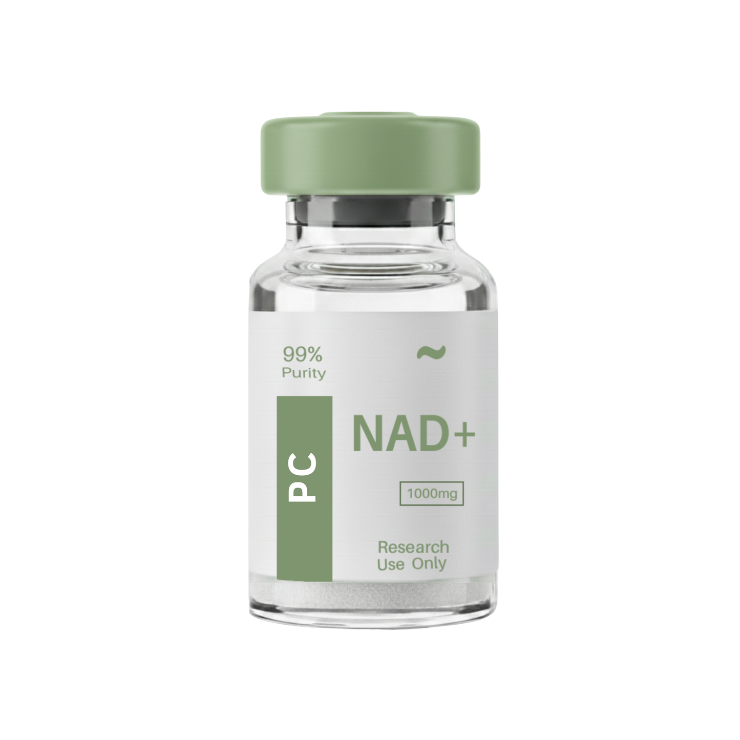
This batch of NAD+ (Nicotinamide Adenine Dinucleotide) Peptide has been third party lab tested and verified for quality.
Contents: NAD+
Form: Powder
Purity: 99.6%
View Third-Party Tests from Our Partners:

OFF
Couldn't load pickup availability
This product is Shipped & Tested in Canada.
Shipping & Delivery
We are committed to delivering fast, reliable, and transparent shipping for all orders. Please review our policy below for details on delivery times, tracking, and what to expect with every purchase.
View Full Shipping Policy


Proof, not promises.
Third-Party Tested
Independent third-party labs confirm identity and purity on every single lot — not just batch samples.
≥99% Purity
Measured, documented, and published - not claimed. Our certificates of analysis are available for every product.
Lot Traceable
Every COA number matches the exact vial in your hands. Full traceability from lab to your door.
Tracked & Fast Shipping
All orders ship with full tracking so you always know where your package is. Fast delivery on every order.
You may also like
-
5-Amino-1MQ
Regular price $80.00Regular price $80.00 Sale priceUnit price per$105.0023% -
Acetic Acid Water 0.6%
Regular price From $14.00Regular price From $14.00 Sale priceUnit price per$19.0026% -
Benzyl Alcohol 0.9%
Regular price From $14.00Regular price From $14.00 Sale priceUnit price per$19.0026% -
BPC-157 + TB-500
Regular price From $97.00Regular price From $97.00 Sale priceUnit price per$127.0023% -
BPC-157 + TB-500 + GHK-Cu
Regular price $155.00Regular price $155.00 Sale priceUnit price per$202.0023% -
Cagrilintide
Regular price From $100.00Regular price From $100.00 Sale priceUnit price per$132.0024% -
Cagrilintide + Semaglutide
Regular price From $125.00Regular price From $125.00 Sale priceUnit price per$181.0030% -
Cerebrolysin
Regular price $40.00Regular price $40.00 Sale priceUnit price per$54.0025% -
CJC-1295 (No DAC)
Regular price From $75.00Regular price From $75.00 Sale priceUnit price per$98.0023% -
CJC-1295 No DAC & Ipamorelin
Regular price From $59.00Regular price From $59.00 Sale priceUnit price per$89.0033% -
CJC-1295 with DAC
Regular price $139.00Regular price $139.00 Sale priceUnit price per$182.0023% -
Epitalon (Epithalon)
Regular price From $50.00Regular price From $50.00 Sale priceUnit price per$67.0025% -
Glow BPC-157 + GHK-CU + TB-500
Regular price $139.00Regular price $139.00 Sale priceUnit price per$181.0023% -
Glutathione
Regular price $83.00Regular price $83.00 Sale priceUnit price per$109.0023% -
HGH 191AA (Somatropin)
Regular price From $55.00Regular price From $55.00 Sale priceUnit price per$72.0023% -
HGH Fragment 176-191
Regular price $97.00Regular price $97.00 Sale priceUnit price per$127.0023% -
Hyaluronic
Regular price $28.00Regular price $28.00 Sale priceUnit price per$37.0024% -
IGF-1 LR3 (Long R3)
Regular price From $40.00Regular price From $40.00 Sale priceUnit price per$53.0024% -
Ipamorelin
Regular price From $32.00Regular price From $32.00 Sale priceUnit price per$42.0023% -
Kisspeptin-10
Regular price From $65.00Regular price From $65.00 Sale priceUnit price per$85.0023% -
KLOW Blend - GHK-CU + TB-500 + BPC-157 + KPV 10mg
Regular price $200.00Regular price $200.00 Sale priceUnit price per$261.0023% -
KPV Tripeptide
Regular price From $56.00Regular price From $56.00 Sale priceUnit price per$74.0024% -
L-Carnitine
Regular price $97.00Regular price $97.00 Sale priceUnit price per$127.0023% -
Lemon Bottle 10mg
Regular price $80.00Regular price $80.00 Sale priceUnit price per$105.0023% -
Lipo-C with B Vitamins
Regular price $85.00Regular price $85.00 Sale priceUnit price per$112.0024% -
Melanotan II (MT2)
Regular price $50.00Regular price $50.00 Sale priceUnit price per$67.0025% -
Oxytocin Acetate
Regular price $42.00Regular price $42.00 Sale priceUnit price per$57.0026% -
Reconstitution Solution
Regular price $15.00Regular price $15.00 Sale priceUnit price per$20.0025% -
Retatrutide
Regular price From $69.00Regular price From $69.00 Sale priceUnit price per$118.0041% -
Semaglutide
Regular price From $36.00Regular price From $36.00 Sale priceUnit price per$47.0023% -
Sermorelin
Regular price From $70.00Regular price From $70.00 Sale priceUnit price per$92.0023% -
SLU-PP-332
Regular price $125.00Regular price $125.00 Sale priceUnit price per$164.0023%


Frequently Asked Questions
Here you'll find answers to common questions.
Every vial we sell comes from a lab that follows current Good Manufacturing Practices (cGMP). That means each step of production is documented and controlled. Before a batch is released, it’s tested by independent third-party labs for purity, identity, and sterility. Certificates of analysis are available so you can see the exact test results.
Yes. The labs we work with use ISO-certified clean rooms where air quality, equipment, and handling procedures are tightly regulated. Staff are trained to pharmaceutical-grade standards. This ensures the peptides are produced in an environment that minimizes contamination risks.
Peptides in lyophilized (freeze-dried) form are stable at room temperature for transport. Once you receive them, refrigeration is recommended to maintain long-term integrity. We package every order securely to prevent damage and ship promptly, so your vials arrive in optimal condition.
We operate under strict in-house protocols that follow current Good Manufacturing Practices (cGMP). That means our team oversees the entire process from sourcing raw amino acids to the final lyophilized vial. Nothing is outsourced or repackaged. This gives us full control over purity, consistency, and sterility, and it’s why we can stand behind every single vial we ship.
Store them in the refrigerator, away from direct light and heat. If you need to keep them longer, some peptides can be stored frozen. Each vial comes with clear handling instructions so you know the proper conditions for stability.
The strongest proof is transparency. For every peptide, we can provide certificates of analysis, manufacturing documentation, and references to the published scientific research behind it. If you ever have questions, we’ll show you the data rather than ask you to take our word for it.
The difference is transparency. Most sites give you a product name and a price. We provide full batch testing, lab documentation, and direct access to certificates of analysis so you don’t have to guess what you’re getting. When you order from us, you know exactly what’s in the vial, where it was made, and how it was verified.
What Our Customers Say
"Absolutely worth it."
Best decision I made all year. Product arrived fast, purity was confirmed, and the results in my research have been consistent from batch to batch. Nothing comes close at this price point.
"Legit. Period."
Ordered twice now. Both times the COA matched exactly what was advertised. You can actually verify everything they claim. That level of transparency is almost unheard of in this space.
"Pure. Verified. Trusted."
I have been sourcing peptides for research for four years. Windsor Peptides is the first company that has made me feel completely confident in what I am ordering. The documentation is real, the purity is real, and the customer service team actually knows what they are talking about when you call.
"Solid every time."
Four orders in and I have had zero complaints. Consistent purity, fast shipping, and the lab reports are always current. I knocked one star only because I wish the website had more product info. Otherwise flawless.
"Best source available."
Switched from a competitor six months ago and the difference was immediate. Better reconstitution, cleaner results, and actual proof of purity. I will not go back.
"Unmatched transparency."
Every single claim they make on their website is backed by actual lab documentation. I reviewed the HPLC reports myself and the numbers are exactly what they advertise. As a biochemist I can say that this level of honesty is almost impossible to find in this market.
"Ships fast. Works."
Ordered on a Monday, received Thursday. Vial was sealed and cold. Lab report included. Results speak for themselves.
"Real COA. Real purity."
I was burned by another supplier last year who claimed 99 percent purity but had no documentation to back it up. Windsor Peptides is completely different. You get a real certificate of analysis with every order, tied to the exact lot number on your vial. This is how all companies should operate.
"Exceeded expectations."
Quality is genuinely exceptional. Every batch I have ordered has tested clean and performed exactly as the research literature suggests it should. I recommend Windsor Peptides to every researcher in my network.
"Finally, real quality."
After trying four different suppliers over two years and being consistently disappointed, I finally found a source I can trust. Third-party verified purity, detailed COA, and a team that responds quickly to technical questions.
"Worth every penny."
Honestly could not be happier. The purity is verified, the shipping was fast and discreet, and the product performed exactly as expected in my protocols. Will be a repeat customer for as long as they are operating.
"Legit. No doubt."
I reviewed the certificate of analysis from the third-party lab myself. The numbers are real. The purity numbers match. The molecular weight is confirmed. This is the only supplier I have found that can back every single claim with actual documentation.
"Simply the best."
Consistent. Verified. Fast. I have recommended them to every colleague in my lab and they have all had the same positive experience I have had since day one.
"Go-to supplier."
I have placed over twenty orders. Not once has the quality been anything other than exceptional. The third-party COA documentation is always available and always accurate. This is my permanent go-to for all peptide research needs.
"Impressive documentation."
The COA packet I received with my first order was more detailed than anything I had seen from other suppliers. HPLC chromatogram, mass spec confirmation, endotoxin test, and sterility results all included. This is what research-grade actually means.
"No hesitation."
Shipment arrived within four days. Packaging was professional and cold on arrival. The included lab report matched the lot number on the vial. Everything about this company says they take quality seriously.
"Consistent. Every order."
Ten orders over the past two years and not a single one has disappointed me. The purity is always verified, the shipping is always fast, and the team always answers questions accurately. I trust this company completely.
"Nothing compares."
I have spent years trying to find a reliable, transparent peptide supplier. After going through six different companies, I found Windsor Peptides three years ago and have never looked anywhere else since. The COA is genuine, the purity is real, and the service is excellent.
"Top tier quality."
The peptides perform exactly as described in the published literature. That alignment only happens when purity is actually as claimed. Windsor Peptides delivers on every promise and backs it with verifiable documentation.
"Earned my trust."
Trust in this industry is rare. Windsor Peptides earned mine through consistent product quality, honest documentation, and a support team that actually understands what they are selling. Three years as a customer and I have never been let down once.
"Absolutely worth it."
Best decision I made all year. Product arrived fast, purity was confirmed, and the results in my research have been consistent from batch to batch. Nothing comes close at this price point.
"Legit. Period."
Ordered twice now. Both times the COA matched exactly what was advertised. You can actually verify everything they claim. That level of transparency is almost unheard of in this space.
"Pure. Verified. Trusted."
I have been sourcing peptides for research for four years. Windsor Peptides is the first company that has made me feel completely confident in what I am ordering. The documentation is real, the purity is real, and the customer service team actually knows what they are talking about when you call.
"Solid every time."
Four orders in and I have had zero complaints. Consistent purity, fast shipping, and the lab reports are always current.
"Best source available."
Switched from a competitor six months ago and the difference was immediate. Better reconstitution, cleaner results, and actual proof of purity. I will not go back.
"Ships fast. Works."
Ordered on a Monday, received Thursday. Vial was sealed and cold. Lab report included. Results speak for themselves.
"Go-to supplier."
I have placed over twenty orders. Not once has the quality been anything other than exceptional. The COA documentation is always accurate. This is my permanent go-to for all peptide research needs.
"Earned my trust."
Trust in this industry is rare. Windsor Peptides earned mine through consistent product quality, honest documentation, and a support team that actually understands what they are selling. Three years as a customer and I have never been let down once.
"Top tier quality."
The peptides perform exactly as described in the published literature. That alignment only happens when purity is actually as claimed. Windsor Peptides delivers on every promise and backs it with verifiable documentation.
"Nothing compares."
I have spent years trying to find a reliable peptide supplier. After going through six different companies, I found Windsor Peptides three years ago and have never looked anywhere else since.
"Premium quality guaranteed."
I specifically chose Windsor Peptides because of their commitment to third-party testing. The COA I received with my last order showed 99.4 percent purity confirmed by HPLC. That is not a claim — that is a documented result from an independent lab. Remarkable standard.
"Verified. Reliable. Fast."
Every order shows up on time, every COA is accurate, every product performs as expected in my lab. I have recommended this supplier to my entire research team and every one of them has had the same reliable experience.
"Lab-verified. No excuses."
The peptide industry has a serious problem with unverified quality claims. Windsor Peptides solves that problem by providing real COA documentation for every single product. You can look up the batch number and verify the results yourself. I cannot recommend them enough.
"Fast. Clean. Done."
Ordered Wednesday, arrived Friday. Cold-packed, sealed, complete certificate of analysis included. Exactly what you expect from a premium supplier. I have reordered three times now without hesitation.
"Outstanding. Full stop."
I have been in research for fifteen years. I know what good sourcing looks like. Windsor Peptides meets every standard I apply when evaluating a supplier. Their documentation is thorough, their purity is verified, and their team is genuinely knowledgeable.
"Highly recommend!"
Clean product, verified results, and a supplier that actually stands behind what they sell. I researched a dozen companies before choosing Windsor Peptides and it was obviously the right call.
"Unreal purity levels."
I have independently tested products from five different suppliers. Windsor Peptides consistently delivers the highest purity levels of any source I have used. Their commitment to third-party verification is not marketing — it is a documented commitment they keep on every single order.
"Very reliable source."
Seven orders over the past year and a half. Delivery averages three to four days, the purity documentation is always included, and the product always performs as expected. Minor packaging improvement would make this five stars but quality is genuinely excellent.
"Game changer."
Switching to Windsor Peptides completely changed how I approach sourcing for my research. I no longer have to wonder if what I received matches what was advertised. The documentation answers every question before I even ask it.
"Premium without compromise."
Ordered my first batch skeptically — I had been burned before by suppliers with impressive websites but terrible products. Windsor Peptides was the opposite. The documentation was thorough, the purity was verified, and the results in my protocols were immediately better than anything I had been getting before.
"Exactly as advertised."
Every product I have ordered has arrived exactly as described. The purity matches the documentation. The results match the research literature. That consistency over multiple orders is how you know a supplier is genuinely committed to quality.
"True professionals."
Everything about Windsor Peptides says professional-grade operation. The documentation, the packaging, the responsiveness of support, and above all the product itself. I use no other supplier and have no intention of changing that.
"Industry standard setter."
If every peptide company operated the way Windsor Peptides does, this industry would have a completely different reputation. Independent testing on every lot, full COA available, and a team that treats you like a professional.
"Repeat customer. Always."
I found Windsor Peptides two years ago after endless frustration with unreliable suppliers. Since then I have placed fourteen orders without a single issue. Simply the best peptide source available.
"No one else compares."
I have tried eight different suppliers. The only consistent, fully-documented, third-party verified experience I have ever had is with Windsor Peptides. Everyone else either lacked documentation, had inconsistent purity, or simply could not match the quality standard this company maintains on every order.
"Lab-perfect results."
The peptides perform in my protocols exactly the way the published research says they should. That only happens when the purity is genuinely as high as the supplier claims. Windsor Peptides delivers on that promise without exception.
"Flawless experience."
From ordering to delivery to results — flawless. I have been researching peptides for six years and Windsor Peptides has become my only trusted source. Their commitment to verified, documented purity is something I have found nowhere else at this price point.
"Gold standard supplier."
Third-party verified on every lot, fast shipping from Canada, and a customer service team that actually understands peptide science. This is what the gold standard of peptide sourcing looks like.
"Always impressed."
Every order impresses me. The purity documentation is thorough, the product quality is consistent, and the support team is knowledgeable and responsive. I have been a customer for two and a half years and my confidence in this company has only grown over time.
"Simply outstanding."
Five years of research, dozens of orders, zero disappointments. The level of transparency Windsor Peptides brings to this market is unmatched. Real documentation, real purity, real results every single time.